Milia
OVERVIEW
What are milia?
Milia, commonly known as "fat granules," also referred to as white acne or milia lichenoides, are tiny subepidermal keratin cysts measuring only 1–2 mm in size [1].
Spontaneous milia are small epidermoid cysts derived from the infundibulum of vellus hair (the fine hair on the body, excluding scalp, armpit, and pubic hair, commonly known as "peach fuzz"). They can also occur secondary to superficial trauma, medication, or other skin conditions due to keratin clogging the eccrine ducts or hair follicles [1].
Milia in infants usually resolve spontaneously within about a month, whereas lesions in adults may persist for a long time.
Is milia a genetic disease? Is it serious?
The onset of some primary milia is influenced by genetic factors and follows an autosomal dominant inheritance pattern [2].
This condition is benign and not serious. It typically causes no symptoms such as pain or itching and does not require treatment unless for cosmetic concerns.
Is milia common?
Milia is extremely common and can occur in individuals of any age or gender. Up to 50% of newborns, regardless of race or sex, develop milia at birth or shortly after, lasting for several weeks [3]. In other words, up to 50 out of 100 newborns will develop milia at birth or shortly thereafter.
Which department should I visit for milia?
Dermatology.
SYMPTOMS
What are the types of milia, and what are their manifestations?
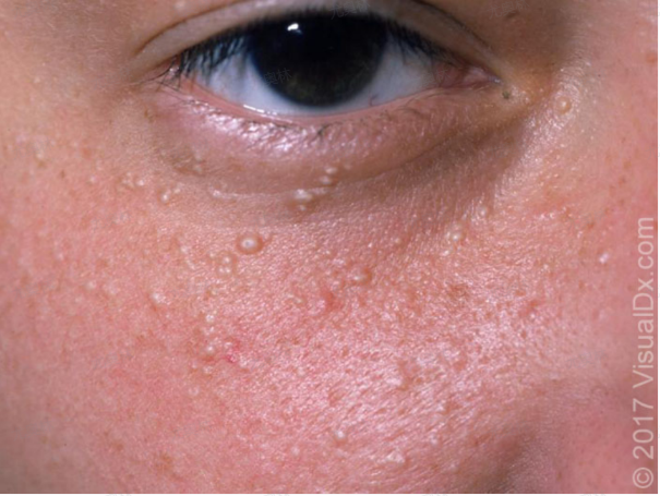
Milia can be divided into primary and secondary types. The rash is characterized by white, tiny subepidermal keratin cysts, about 1–2 mm in size, mostly round, resembling small white sesame seeds. Patients typically experience no special sensations, with no pain or itching symptoms [1]. When punctured, a sebum-like substance can be squeezed out, hence the term "fat granules," though the extracted material is actually fragments of keratin.
- Primary milia: Often occurs spontaneously, commonly on the face, especially the eyelids, nose, scalp, upper trunk, and proximal limbs. It can occur at any age or gender but is most frequent in newborns. In infants, milia may appear around the areola and persist. In adults, it is more common in young women and can also occur on the genitals. Primary milia has no clear cause, though some cases may involve genetic factors.
- Secondary milia: Often found on the earlobes, back of the hands, forearms, or areas of trauma. It may also result from adverse drug reactions or secondary to certain skin conditions involving epidermal damage (e.g., epidermolysis bullosa, porphyria cutanea tarda, bullous lichen planus, dermatitis herpetiformis, pemphigus, or bullous pemphigoid).
Why is milia commonly seen around the eyes?
The most noticeable feature of the skin around the eyes is its thinness—only about one-third the thickness of facial skin.
Additionally, the eye area has poor circulation, slow metabolism, and weak absorption, making it a common site for milia and other skin conditions like syringomas, flat warts, and hemangiomas.
Does milia on a newborn's face affect their health?
No, it does not.
Newborn skin is very thin, and about 50% of infants develop milia, either at birth or shortly after. This means that out of 100 newborns, 50 may have milia. These milia appear as dense, tiny clusters with no obvious granular texture [3].
Mothers need not worry—these milia will disappear naturally as the baby's metabolism progresses and do not affect the baby's health.
What are the manifestations of milia during pregnancy?
The most common area for milia during pregnancy is the face, particularly around the eyes. The typical appearance is small, whitish, firm bumps, mostly 1–2 mm in diameter and rarely exceeding 3 mm.
These bumps may appear singly or in clusters and usually persist without significant changes in size over a short period. They cause no itching or pain.
CAUSES
What causes milia?
- Primary causes: Can occur in newborns or individuals of any age or gender. It is formed by undeveloped sebaceous glands, disordered fat metabolism, or due to endocrine imbalances and low skin regulation ability. The lesions may disappear naturally within 1–3 months [4].
- Secondary causes: May develop after procedures like dermabrasion, abrasions, scratching, sun exposure, second-degree burns, certain skin conditions (e.g., epidermolysis bullosa, porphyria cutanea tarda, bullous lichen planus, dermatitis herpetiformis, pemphigus, bullous pemphigoid), or X-ray exposure [5].
- Improper use of topical medications or cosmetics: Long-term use of corticosteroid ointments can trigger milia. Excessive use of heavy eye makeup, exfoliants, or scrubs may cause microscopic, invisible wounds around the eyes, leading to milia during skin repair. Overly oily cosmetics can clog hair follicles and induce milia.
- Genetic factors: Some cases are hereditary. The condition follows an autosomal dominant pattern, meaning if one parent carries the dominant gene for milia, the fetus may inherit the condition [2].
Who is more prone to milia?
- People who wear makeup: Inadequate skin cleansing may increase the risk.
- Outdoor workers: Prolonged exposure to UV radiation raises the likelihood of developing milia.
- Those with a family history: Since genetics play a role, individuals with affected relatives have a higher risk.
Why do milia appear during pregnancy?
Milia can develop at any stage of life, and their occurrence during pregnancy is not directly related to gestation.
The main causes fall into two categories:
- Spontaneous formation (primary milia):
No obvious cause; milia appear without a clear trigger.
- Secondary factors:
Milia may arise after skin trauma (e.g., cuts, scratches, burns, blisters, abrasions, laser treatments) or other skin conditions.
Despite being colloquially called "fat granules," milia have no connection to obesity or excessive nutrition.
DIAGNOSIS
How is milia diagnosed?
The disease can be diagnosed based on typical rash morphology combined with dermatoscopy. Generally, no special tests are required. When the skin lesions are atypical and visual diagnosis is inconclusive, a histopathological examination may be performed.
What tests are needed for milia?
- Physical examination: Preliminary diagnosis can be made by visual inspection of the appearance and location of the skin lesions.
- Dermatoscopy: This non-invasive method can reveal morphological features not visible to the naked eye, aiding in diagnosis. Under dermatoscopy, milia appear as bright white homogeneous structures [4].
- Histopathological examination: The pathological characteristics of milia help confirm the diagnosis and differentiate it from other conditions. The typical finding is "a stratified squamous epithelial cyst wall with a granular layer and laminated keratinous contents" [1].
How to distinguish milia from acne?
Milia typically appear as small, white or yellow granular bumps, resembling acne. The key difference is that whiteheads in acne can be easily squeezed out by hand, whereas milia, covered by keratin, are stubborn and difficult to extract.
How to distinguish milia from syringoma?
Both syringoma and milia commonly occur around the eyes. Milia are small, round, and uniform in shape, appearing white or pale yellow. Syringoma, however, are irregularly shaped, often resembling commas or tadpoles, and are light brown. Syringoma requires laser treatment for removal.
Syringoma is associated with endocrine factors, pregnancy, menstruation, and genetic predisposition, primarily affecting adolescent females. Milia, linked to endocrine factors, genetics, skin conditions, and sun exposure, can occur at any age or gender but are most common in newborns.
TREATMENT
How to Treat Milia?
In fact, no treatment is necessary as it generally does not affect health, and some may resolve on their own. If you find it aesthetically bothersome and wish to remove it, you can take any of the following measures:
1. Needle Extraction:
It is best to visit a dermatology department at a hospital for this procedure. Self-treatment is not recommended to avoid accidental injury to the eye area or even the eyes.
The general process is as follows:
- First, sterilize the specialized needle, then disinfect the milia area around the eyes with iodine solution.
- Use the sterilized needle to pierce the milia to a certain depth and gently lift the epidermis.
- Then, use two sterilized cotton swabs or a comedone extractor to press the base of the milia to expel the secretions inside.
- Afterward, disinfect again with iodine solution or alcohol, and avoid wetting the wound for 24–48 hours to prevent infection.
Generally, milia should only be extracted when they are mature (i.e., when they turn white or pale yellow). If they recur, consult a professional for another extraction. The downside is that improper handling or care during the procedure may leave scars.
2. Laser Treatment:
To significantly reduce the chance of recurrence, laser treatment [1] can be used. Usually, one session is sufficient, but if it recurs, additional treatment may be needed.
The cost depends on the number of milia, typically charged per lesion (around a few dozen yuan each), with regional price variations.
The general process is as follows:
- First, disinfect the milia and surrounding skin with iodine solution.
- Apply local anesthesia (immediate effect) or topical anesthetic (takes about 30 minutes to work).
- Use a laser device, such as a CO₂ laser, to completely remove and destroy the milia.
- Disinfect again with iodine solution and keep the wound dry for 3–5 days to prevent infection.
Laser treatment can thoroughly remove the lesions, is fast, and rarely leaves scars. However, larger papules may still scar.
3. Topical Medications:
Topical medications, such as tretinoin cream, can be used. This method is simple, non-invasive, and inexpensive, but due to its lower efficacy compared to needle extraction or laser, it is less commonly used clinically. Note the following:
- These medications work slowly and require consistent use to improve milia.
- The drugs can be irritating, so gradual tolerance building is necessary (follow medical advice for proper usage to allow the skin to adapt).
- This treatment is not suitable during pregnancy, breastfeeding, or when trying to conceive.
Additionally, maintain facial cleanliness, avoid excessive exfoliation or heavy makeup, and choose lightweight eye creams and skincare products.

How to Treat Milia During Pregnancy?
Milia itself is neither painful nor itchy and does not affect health, so treatment is not mandatory.
If you find it aesthetically bothersome or emotionally distressing, removal is an option. The general recommendation is physical treatment, such as making a small incision with a blade and using a comedone extractor to remove the accumulated keratin.
This can be done during pregnancy, with recovery taking just a few days and minimal scarring. However, even though the procedure is simple, self-treatment at home is not advisable due to risks of inadequate sterilization or improper technique.
Since milia is a localized skin structural issue, topical medications have limited effectiveness and are not highly recommended.
DIET & LIFESTYLE
What should patients with milia pay attention to in daily life and diet?
- After needle extraction or laser treatment, maintain wound hygiene. Let scabs fall off naturally and avoid scratching the affected area to prevent infection.
- Engage in moderate exercise (e.g., jogging, walking) to improve physical fitness, but avoid prolonged sun exposure during workouts.
- No specific dietary restrictions are required. Maintain a balanced and nutritious diet.
Does milia require follow-up examinations?
Yes. After laser treatment, patients should return for a check-up within 1-3 months. For needle extraction, follow-up is needed within 1-2 weeks post-treatment.
Is milia related to eye cream usage?
When milia appears, people often blame it on "overly rich eye cream!" In fact, this is a misconception!
Some individuals habitually use heavy eye makeup or excessive exfoliants, which may cause milia. Since they often choose rich eye creams for dry skin, they mistakenly attribute milia to the cream.
How to choose eye creams and cosmetics for milia?
Although no scientific evidence proves cosmetics directly cause milia, skin irritation from cosmetic ingredients can lead to secondary milia.
When selecting skincare products, prioritize non-irritating ingredients and allergen-free formulas to minimize skin damage.
Key selection criteria: low irritation (free of alcohol, preservatives, mineral oil), and suitability for your skin type and age.
How should pregnant women care for milia?
Milia doesn't affect health and requires no special care.
Daily recommendations: Reduce frequent skin friction, protect skin, and avoid injuries.
No specific precautions are needed for diet, sleep, or skincare.
PREVENTION
How to Prevent Milia?
- Keeping the eye area clean is most important. Use a cleanser correctly and moderately (squeeze the cleanser into your palms, rub to create foam, apply to the face—no more than twice a day). If the skin is not thoroughly cleansed in time, pores may become clogged, preventing excess oil from being expelled, leading to raised milia.
- Choose low-irritation skincare products (free of alcohol, mineral oil, etc.) that are suitable for your age.
- Pay attention to your massage technique. While various anti-aging or absorption-boosting massage methods abound, ensure the pressure and positioning are correct (use your index, middle, and ring fingers to gently massage along the lower eye socket from the inner to outer corner; then place your thumb at the inner corner and lightly massage the upper eyelid outward, finishing with gentle pressure on the temples). Incorrect techniques may damage delicate eye-area skin, causing wounds that trigger milia.
How to tell if the massage is too harsh? A slight stinging sensation indicates skin damage—stop immediately.