Fetal ventricular echogenic focus
What is a fetal ventricular echogenic focus? What causes it?
Commonly seen in mid-pregnancy ultrasound examinations
Fetal ventricular echogenic focus, also known as echogenic intracardiac focus (EIF), is typically detected during mid-pregnancy ultrasound examinations (mostly between 18–27 weeks of gestation).
It appears as a punctate or patchy hyperechoic area in the ventricular chordae (a structure that anchors the heart valves) or papillary muscles (which connect to the ventricular wall and extend chordae to secure the valves), moving synchronously with the heartbeat. See the image below.
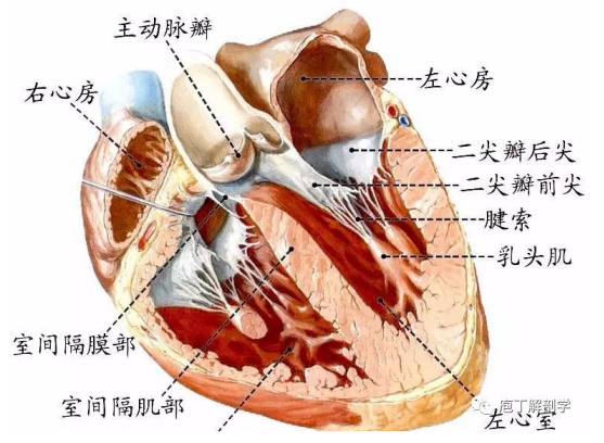
Image source: Internet
On ultrasound, a fetal ventricular echogenic focus appears as a bright, white punctate or patchy image without acoustic shadowing, similar to a dark shadow, usually measuring 1–6 mm in diameter.
Generally, the incidence of fetal ventricular echogenic foci in mid-pregnancy is about 0.13%–20% [1], averaging around 4.5% [2]. Most cases occur in the left ventricle, with a few in both ventricles or the right ventricle, and they may appear as single or multiple foci.
It is a very common clinical finding, with approximately 95% of ventricular echogenic foci resolving naturally by late pregnancy.
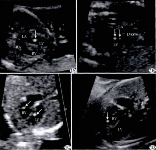
Image source: Internet
Exact cause remains unclear
The exact cause of fetal ventricular echogenic foci is still unknown but may be related to thickening of the ventricular chordae, calcification or mineral deposition in the papillary muscles, or incomplete perforation of the papillary muscles and chordae [3].
Additionally, fetal ventricular echogenic foci may be associated with ethnicity, with higher prevalence rates observed in Asian populations [4-6].
This condition may also be linked to pathological factors such as congenital heart disease (e.g., ventricular septal defect, tetralogy of Fallot, pulmonary valve stenosis, hypoplastic left heart syndrome) or chromosomal abnormalities, though no definitive conclusions have been reached.
What should you do if a fetal ventricular echogenic focus is detected on ultrasound?
No need to worry excessively; most cases are normal
It is a very common clinical phenomenon, and the vast majority of ventricular echogenic foci do not cause cardiac abnormalities. They are often unrelated variations during ventricular development and generally do not adversely affect fetal growth. About 95% of these foci resolve naturally by late pregnancy.
Follow medical advice for fetal echocardiography
Typically, when a fetal ventricular echogenic focus is detected, doctors will recommend a fetal echocardiogram to check for coexisting structural cardiovascular anomalies or cardiac abnormalities.
If the echocardiogram shows no issues, the fetal ventricular echogenic focus is likely a transient benign change. Expectant mothers need not be overly concerned, and there are currently no specific medications or methods to reduce or eliminate it. Regular follow-up examinations are sufficient.
Conversely, if the fetal echocardiogram reveals other structural or cardiovascular problems, further management—such as consultation with a cardiothoracic surgeon or termination of pregnancy—may be required based on the specific situation.